Echocardiography (echo) is an ultrasound test examining the heart’s structure and function. By types of echocardiography, a multi-dimensional picture of the heart muscle in action – pumping blood into and out of its chambers can be produced. The process is achieved without any blood extraction, piercing, or incisions on the skin, making echo a preferable choice for practitioners and patients.
Echocardiography is versatile. For a patient with symptoms including shortness of breath, pain originating on the left side of the chest, excessive sweating, palpitations, and performing routine tasks, heart echocardiography is the most advisable to assess the condition of the heart. Now please scroll down for more information regarding echocardiography.

What does an Echocardiography Show?
Echocardiography is an ultrasound scan in which a small probe emits high-frequency sound waves that bounce off different body parts and produce echoes. During the scan, these echoes are retrieved by the probe and then converted into a moving image on the monitor.
In line with the images, professionally trained healthcare professionals can explicate:
- Changes in the Heart Size
Echocardiography can show changes in the chambers’ size and the muscle’s thickness.
A weakened or damaged heart valve, high blood pressure, or other diseases can lead to enlargement of the ventricles or abnormal thickening of the heart walls.
- Pumping Strength
Echocardiography is used to determine EF or Ejection fraction (calculated percentage of blood pumped from a filled ventricle every time the heart beats) and Cardiac Output (Volume of pumped blood in each minute by heart) of the heart. Both these values depict the pumping strength of cardiac muscle.
The inability of the heart to pump sufficient blood to satisfy the body’s needs can lead to signs of heart failure.
- Damage to Heart Muscles
Echocardiography can assist the doctor in determining whether all of the parts of the heart wall are properly functioning and contributing to the heart’s pumping activity.
For example, areas of the heart wall that are weakly moving may be damaged in a heart attack or receive too low an amount of oxygen.
- Valve Problems
An echocardiogram enables the doctor to determine whether patients’ heart valves are wide enough open to allow adequate blood flow or fully closed to prevent blood leakage.
- Heart Defects
Echocardiography is a way to detect the heart’s structure abnormalities. For example, defects such as abnormal heart chambers and connection defaults of the heart with major blood vessels, and other complex fetal cardiac defects can be detected using types of echocardiography.
The overall procedure of heart echocardiography is safe and painless. No recorded risks or side effects are associated during and after the procedure.
Techniques of Echocardiography
Several techniques are used to create pictures of the patient’s heart. The optimal technique depends on their specific conditions. These techniques include:
- Two-dimensional Ultrasound
The 2d echo is the most frequently used method. It produces two-dimensional images that appear as ‘slices’ viewed on a computer screen. Traditionally, these slices can be ‘stacked’ to create a three-dimensional structure.
- Three-dimensional Ultrasound
Advances in technology have made three-dimensional imaging more effective and useful. 3D technology allows the doctor to see the parts of the heart from different angles. For example, new 3d technology can show different aspects of the heart more accurately, including how well it is pumping blood.
- Doppler Ultrasound
Doppler ultrasound can estimate blood flow in the blood vessels by bouncing the ultrasound waves off the red blood cells in the circulation. Whereas normal ultrasound only produces images with sound waves but does not show blood flow.
In addition to the three most common detection techniques mentioned above, color doppler ultrasound, strain imaging, and contrast imaging are also included.
Types of Echocardiography
Based on the different techniques, several types of echocardiography are available. Each type of echocardiography has unique benefits in diagnosing and managing heart disease. They include:
- Transthoracic Echocardiography
Transthoracic echocardiography, or TTE, is the most commonly performed echocardiography test and is noninvasive, taking place entirely outside the body.
TTE is used for the diagnosis and studying of Cardiac wall thickness and motion, left and right ventricular function, size of cardiac chambers, valvular diseases, aortic structure, the pressure inside the heart, and its anatomy.
- Transesophageal Echocardiography
Transesophageal echocardiography (TEE) is useful in obtaining a more detailed image of small heart structures such as the posterior cardiac left atrium and atrial appendage, atrial septum, and pulmonary vein.
For obese patients or patients with obstructive pulmonary diseases such as COPD, it is technically hard to obtain ultrasound images through TTE detection. In such conditions, TEE is the procedure of choice. In this echocardiography process, digital images of the heart are obtained by introducing a transducer at the tip of the endoscope through the patient’s stomach and esophagus.
- Exercise Stress Echocardiography
Exercise stress echocardiography is mainly used to check the blood supply to every part of the heart.
It is performed to evaluate and record the heart’s functioning under stress. The procedure includes resting heart echocardiography as a first step. After that, the cardiac oncologist will guide the patient to run on a treadmill, exercise pedal, or bicycle slowly in the first few minutes and gradually faster for about 10-15 minutes.
Applications of Echocardiography
Echocardiography can assist in diagnosing and monitoring certain heart conditions by examining the heart’s structure and the vessels surrounding it, analyzing how blood flows through, and assessing the heart’s pumping chambers.
Echocardiography can help detect the following conditions:
- Heart Attack: A disease affecting the heart or blood vessels.
- Heart Failure: A chronic, progressive disease where the heart muscle cannot pump enough blood to satisfy the body’s demand for blood and oxygen.
- Congenital Heart Disease: Structural defects of the heart or large blood vessels present at birth.
- Heart Valve Problems: Problems affecting one or several of the heart’s four valves.
- Cardiomyopathy: A heart muscle disease where the heart is deprived of the ability to pump blood effectively.
- Endocarditis: A condition where the tissue inside the heart and heart valves becomes inflamed (red and swollen).
Electrocardiography vs. Echocardiography

Although they both monitor the heart, ECG and echocardiography are two different tests. An ECG uses electrodes to detect electrical impulse abnormalities in the heart, while echocardiography uses ultrasound to detect structural abnormalities in the heart. Depending on their condition, a person may need both an ECG and echocardiography.
Let’s find the five significant differences between echocardiography and electrocardiography.
- Method and instruction
During electrocardiography, the doctor or technician mounts 10 small electrodes attached to the machine in different locations on the patient’s chest wall. The sticky pads will record the heart activity through electrical activity and interpret it on a tracing chart. For a small number of male patients, requesting that they shave their chest hair to place the pads correctly and thus obtain excellent tracings may be necessary.
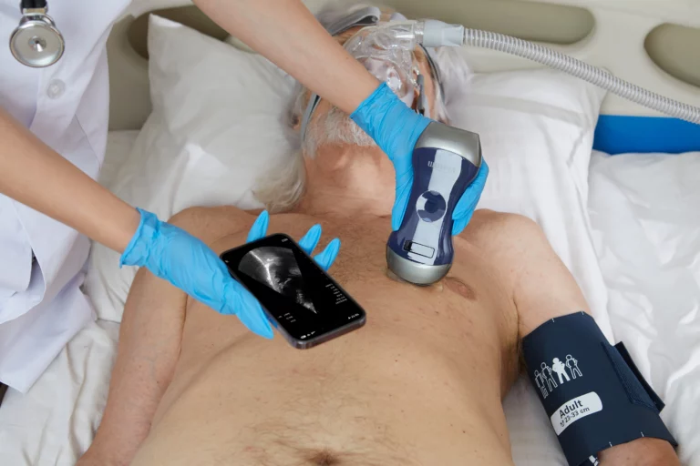
Regarding echocardiography, the patient must lie on a table specially prepared for the examination. In addition, the patient needs to lie on the left side with the left arm near the head. The doctor or technician then applies ultrasound gel to the chest wall, after which an ultrasound probe is placed on the chest to take pictures of the patient’s heart and valve structures. The technician then uses a mobile device, a transducer, above the chest, which is connected to a monitor. An echo test allows the doctor to capture an image of the patient’s heart in the monitor.
- Working principle
Utilizing high-frequency sound waves (ultrasound), echocardiography provides a dynamic image of the patient’s heart. The sound waves are sent through a transducer device into the body. The sound waves then bounce off the heart and return to the transducer in the form of echoes. These echoes are converted into images on a television monitor, producing a picture of the patient’s heart in motion.
The basic principle of electrocardiography lies in the fact that the stimulation of a muscle changes the electrical potential of the muscle fibers. These impulses are transmitted between cells via gap junctions that connect the heart cells. When the cell is at rest, its interior is negatively charged compared to its exterior. Depolarization triggers the heart muscle’s contraction, resulting in a temporary loss of the negative charge inside. However, after depolarisation, the myocardial cells return to their resting charge again, known as repolarization. These depolarisation and repolarisation waves indicate electrical currents and can be monitored by placing electrodes on the surface of the body. After the current has spread from the heart to the body, the ECG picks up these changes and records the activity on previously sensitized paper. An ECG is, therefore, a graphic representation of the electrical activity in the heart. Finally, the current is transmitted over selected electrodes on the ECG machine at the point of contact with the body.
- Availability
Both ECG and echo are noninvasive heart tests and are not painful for the patient. Both are conducted by doctors; nevertheless, an echo test is not as common as an ECG. Basically, every patient who is assessed for a heart-related problem or heart-related symptoms will have an ECG. An ECG can also be considered a screening test and is done on almost everyone with a medical history and physical examination. An echo test is a more specific requirement when there are signs and symptoms of heart disease present.
- Detect
An ECG is an electrical reading, whereas echocardiography is a mechanical reading of the patient’s heart. Consequently, there are differences in detection and findings between echocardiography and electrocardiography.
Generally, An electrocardiography can detect arrhythmias and coronary heart disease, heart attacks, and cardiomyopathy.
Echocardiography detects problems with the pericardium, the large blood vessels inside the heart, and blood clots inside the heart. Beyond this, it also checks for abnormal holes inside the heart.
- Details
Echocardiography presents more accurate details compared to an ECG.
An ECG is an affordable and quick test to monitor the condition of the heart; however, it is not always accurate. An ECG provides limited comprehensive information, more often related to an impending heart attack. Tracking technology shows cardiologists about the peaks and troughs of the heart rhythm on a continuous timeline.
Echocardiography provides insightful and accurate information about a patient’s heart’s overall structure and function. It shows a color video image of the heart, surrounded by veins and blood vessels. The red, blue, and yellow lights represent the numerous ventricles and the outer walls of the heart. In addition, the test offers accurate information about a weakened heartbeat, squeezed, blocked or blocked blood vessels, and overall structural integrity.
How do ECG and Echocardiogram Complement Each Other?
Despite several differences between echocardiography and ECG, they are the most compatible partners. The echocardiography is a detailed and accurate information-gatherer if the ECG is the clue-giver.
An ECG rapidly collects information about a patient’s heart and provides it in a clearly ordered manner. The ECG measures the electrical efficiency of the heart to determine if the pattern is normal, unstable, too slow, or too fast. If the size or shape of the heart is abnormal, this will also be seen. If there are any heart attack symptoms, the patient may be asked to undergo further tests, namely an ultrasound. For instance, if the patient has chest pain and the ECG shows a possible heart attack, blockage, or abnormal tracing, the doctor may order echocardiography, which is a more in-depth and accurate test.
In short, an ECG is a valid first test, and the doctor will be able to see irregularities that would be signs of heart disease. However, an ECG does not accurately assess the heart’s ability to pump blood. If the doctor wants to check how efficiently the heart is pumping blood, they will need to perform an ultrasound on the patient after the ECG.
Viatom Portable Ultrasound

Any dysfunction in the cardiovascular system can badly affect normal routine life and may eventually be fatal. Therefore, keeping knowledge about cardiac health conditions at intervals is necessary. Heart echocardiography can serve the purpose.
At Viatom, we proudly offer our 3-in-1 portable hand-held wireless Ultrasound scanner. It produces HD images by utilizing the latest digital imaging technology. It is designed to simplify the instrument’s functioning and be easy for the operator.
In brief, our 3-in-1 wireless ultrasound scanner can be conducted easily, economically, and ergonomically. If you are interested, please do not hesitate to contact us for more details.
Reference:
https://www.nhlbi.nih.gov/health/heart-tests
https://www.nhlbi.nih.gov/health/heart-valve-diseases/diagnosis